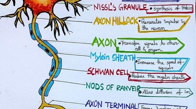
Neuron Anatomy: A Color-Coded ExplorationNeurons are the fundamental units of the brain and nervous system, responsible for receiving sensory input from the external world, sending motor commands to our muscles, and for the intricate operations of the brain, such as thinking, feeling, and perceiving.
Introduction
Neurons are the fundamental units of the brain and nervous system, responsible for receiving sensory input from the external world, sending motor commands to our muscles, and for the intricate operations of the brain, such as thinking, feeling, and perceiving. Neurons come in various shapes and sizes, but they all share a common basic structure. Understanding the anatomy of a neuron is critical to unraveling the complexities of the brain and nervous system. This article will delve into the structure of neurons, color-coded for clarity and ease of understanding, and explore how their various parts work together to carry out their vital functions.
The Structure of a Neuron
A neuron consists of three main parts: the cell body (soma), dendrites, and the axon. Each part plays a distinct and crucial role in the neuron’s ability to communicate with other cells. These components can be understood more easily by using a color-coded image representation, where different colors highlight various parts of the neuron.
1. The Cell Body (Soma)
Color Code: Green
The cell body, or soma, is the neuron’s control center, and it contains the nucleus. The nucleus holds the cell’s genetic material (DNA) and is responsible for regulating the cell’s activities, including its growth and metabolic processes. . The soma integrates incoming signals from the dendrites and decides whether to pass them on. In our color-coded neuron image, the cell body is highlighted in green to emphasize its central role in the neuron’s functioning.
2. Dendrites
Color Code: Blue
Dendrites are the branch-like extensions that protrude from the cell body. Their primary function is to receive electrical and chemical signals from other neurons. Dendrites have numerous receptor sites that are designed to pick up neurotransmitters—chemicals that neurons use to communicate with each other. The signals they receive can be excitatory (promoting the generation of a nerve impulse) or inhibitory (preventing a nerve impulse). By collecting these signals, the dendrites pass the information to the soma for processing. In our color-coded image, dendrites are colored blue to show their critical role in gathering and transmitting signals.
3. The Axon
Color Code: Yellow
The axon is a long, slender projection that extends from the cell body. Its primary function is to transmit the nerve impulses away from the soma to other neurons or to muscles and glands. The axon is insulated by a fatty substance called the myelin sheath (which will be discussed in more detail below), which helps speed up the transmission of electrical signals. In our neuron image, the axon is colored yellow to signify its role as the communication pathway that allows the neuron to transmit messages over long distances.
4. Axon Terminals
Color Code: Red
At the end of the axon are the axon terminals, also known as terminal buttons or synaptic boutons. These small structures are responsible for releasing neurotransmitters into the synaptic cleft, the tiny gap between neurons. The axon terminals are depicted in red in the color-coded image, symbolizing their role in signal transmission and communication between neurons.
Additional Structures: Supporting a Neuron’s Function
These include the myelin sheath, nodes of Ranvier, and synapses, each playing an essential role in how neurons send and receive information.
5. Myelin Sheath
Color Code: White
The myelin sheath is a fatty layer that wraps around the axon of some neurons. The main function of the myelin sheath is to insulate the axon and increase the speed at which electrical impulses travel down the neuron.
6. Nodes of Ranvier
Color Code: Orange
The nodes of Ranvier are small gaps in the myelin sheath that expose the axon. These gaps are essential for the rapid transmission of nerve impulses. Without these nodes, the electrical signal would travel much more slowly. In our neuron diagram, the nodes of Ranvier are color-coded in orange, demonstrating their crucial role in accelerating the nerve signal transmission.
7. Synapse
The Process of Neural Communication: Putting It All Together
- Signal Reception: The process begins when dendrites (colored blue) receive signals from other neurons. These signals can be in the form of neurotransmitters, which bind to receptors on the dendrites.
- Signal Integration: The soma (colored green) receives the signals collected by the dendrites and integrates them.
- Signal Propagation: The action potential travels down the axon (colored yellow). These neurotransmitters cross the synaptic cleft and bind to receptors on the postsynaptic neuron, starting the process again in the next cell.
Conclusion
Through this lens, we gain deeper insights into the nervous system’s remarkable ability to process and respond to information.
